
2020
On June 21, 2022 – July 29, 2022 I did an internship at the University of California in Davis, California, USA.
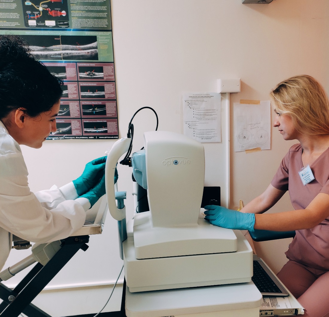 Examination with the use of optical coherence tomography to assess the characteristics of physical and chemical damage to the cornea in domestic pig (Sus domestica).
Comparative Ophthalmic Imaging Laboratory
School of Veterinary Medicine
Uniwersytet Kalifornijski w Davis
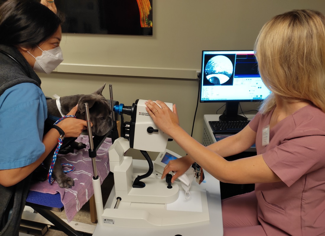 Examination with the use of optical coherence tomography (OCT) of the domestic dog (Canis familiaris).
Comparative Ophthalmic Imaging Laboratory
School of Veterinary Medicine
Uniwersytet Kalifornijski w Davis
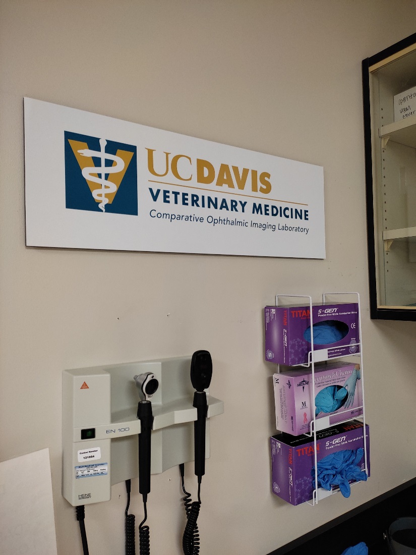 Comparative Ophthalmic Imaging Laboratory
School of Veterinary Medicine
Uniwersytet Kalifornijski w Davis
 School of Veterinary Medicine
Uniwersytet Kalifornijski w Davis
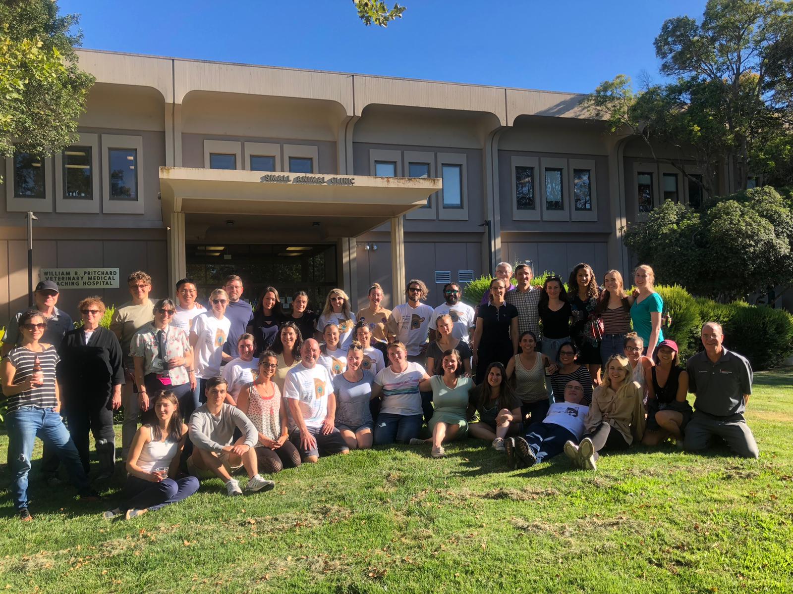 Course participants in front of the building of the University of California Veterinary Hospital in Davis.
16th William Magrane Basic Science Course in Veterinary & Comparative Ophthalmology
School of Veterinary Medicine
Uniwersytet Kalifornijski w Davis
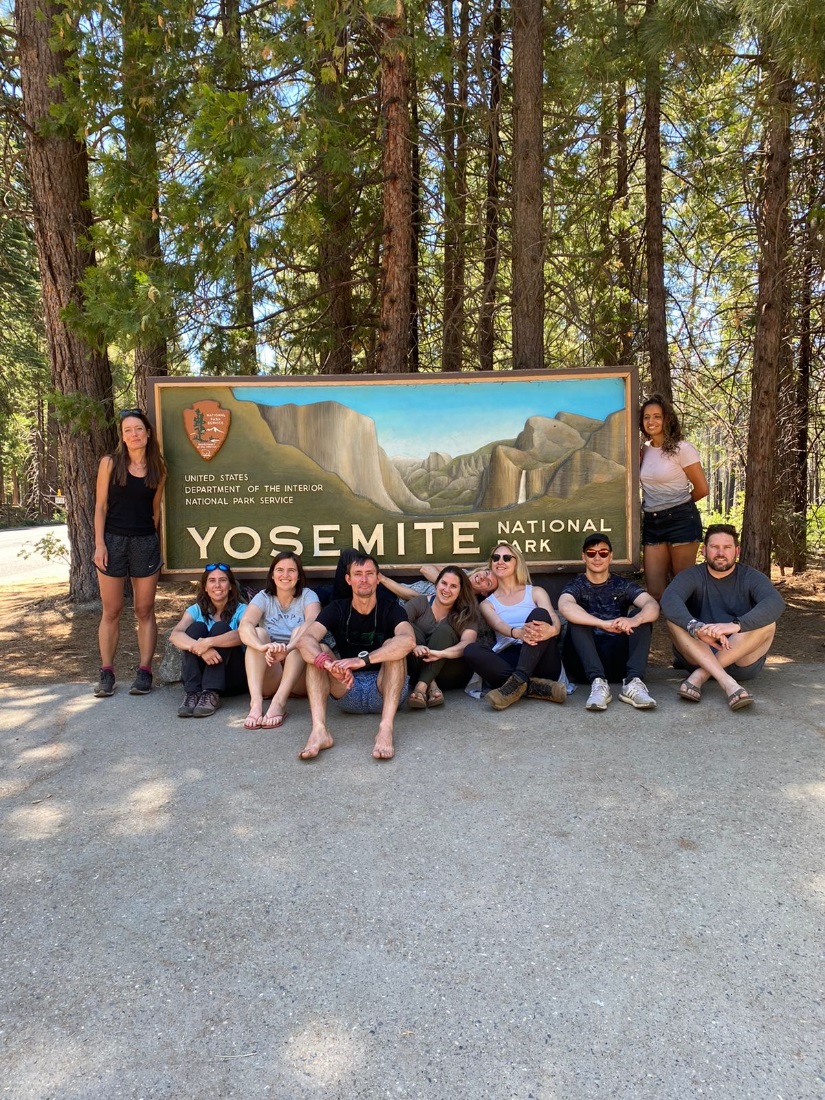 Participants of the 16th William Magrane Basic Science Course in Veterinary & Comparative Ophthalmology.
Park Narodowy Yosemite, Kalifornia, USA
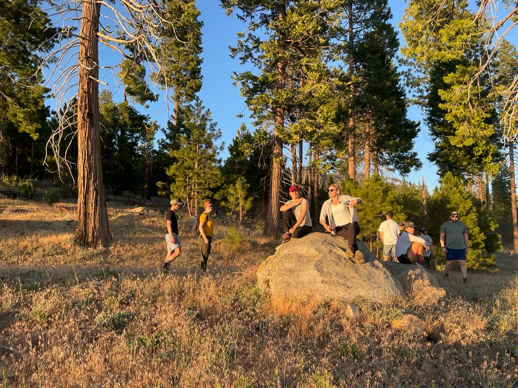 Participants of the 16th William Magrane Basic Science Course in Veterinary & Comparative Ophthalmology.
Park Narodowy Yosemite, Kalifornia, USA
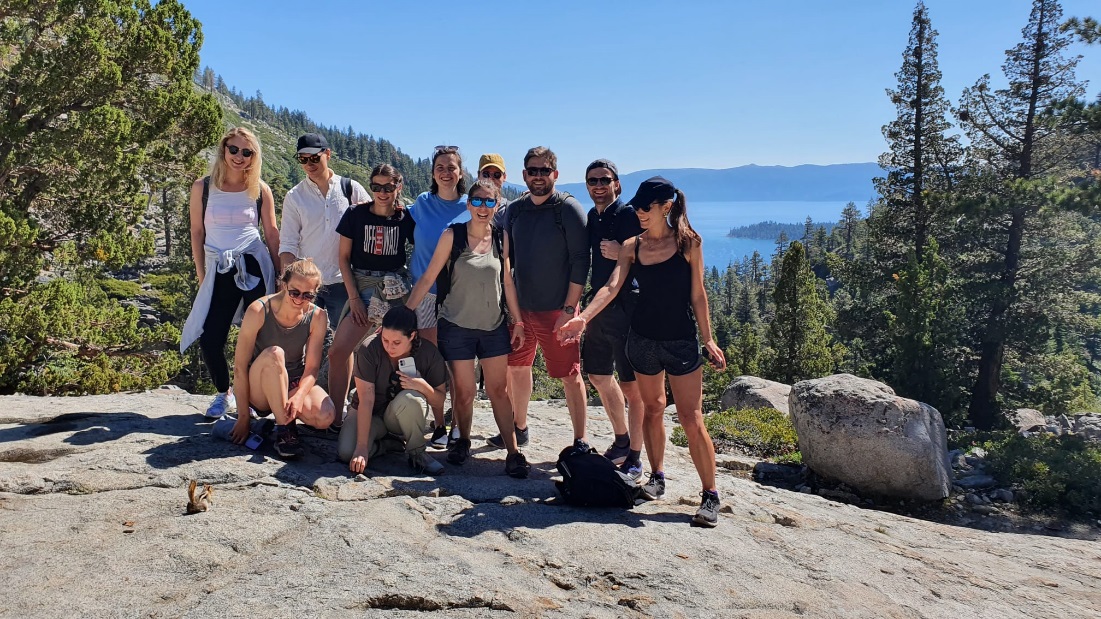 Participants of the 16th William Magrane Basic Science Course in Veterinary & Comparative Ophthalmology
Jezioro Tahoe
Pasmo górskie Sierra Nevada
Kalifornia, USA
The stay at the university consisted of two stages:
1. The first stage was participation in the 16th William Magrane Basic Science Course in Veterinary & Comparative Ophthalmology, attended by 46 veterinarians, incl. from the USA, France, Belgium, South Africa, Spain, Italy, England, Mexico, Sweden, specializing in veterinary ophthalmology. The participants of the course were both clinicians and employees of academic and scientific institutions. Lectures and practical exercises were conducted by leading scientists in the field of veterinary ophthalmology, authors of books and the most important scientific works in this field. The course included both theoretical and practical elements. Participation in the course was extremely valuable in terms of the scope of the latest knowledge in the field of veterinary ophthalmology, as well as in terms of the possibility of establishing foreign cooperation.
2. The second stage of my stay was a research and clinical internship at the University of California Veterinary Hospital in Davis. During the internship, I participated in the clinical activities of the ophthalmology department, both concerning companion, farm and exotic animals. The aim of the clinical internship was to familiarize with the possibilities of using the latest diagnostic devices, including the methods of optical coherent tomography and confocal microscopy, familiarization with the methods of treatment, participation in surgical procedures. The advantage of the internship was the fact that the department of ophthalmology accepted one person for the duration of the internship, which ensured the opportunity to participate in all procedures. During the internship, access to the meetings of residents and professors of the Department of Ophthalmology was also provided, where presentations and discussions were held on the currently published scientific articles in the field of veterinary ophthalmology. I also had the opportunity to participate in didactic activities during student internships and to familiarize myself with the educative methods at the Department of Ophthalmology. Participation in the meetings of professors and residents with students gave me an insight into the practical possibilities of transferring knowledge to trainees. During the internship, I also participated in laboratory and scientific research. Numerous research projects are currently carried out at the Department of Veterinary Ophthalmology. I participated in research with the use of confocal microscopy on the morphological and morphological assessment as well as the corneal endothelial cell density in a domestic dog (Canis familiaris) and changes caused by the phacoemulsification procedure. I also had the opportunity to actively participate in research using the optical method of coherence tomography and histological examination, during which we assessed the characteristics of physical and chemical damage to the cornea in domestic pig (Sus domestica).
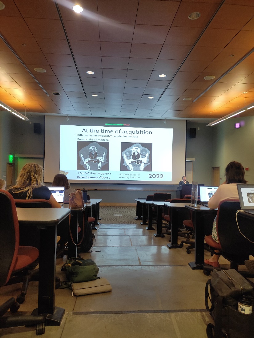 16th William Magrane Basic Science Course in Veterinary & Comparative Ophthalmology
Gladys Valley Hall, UC Davis
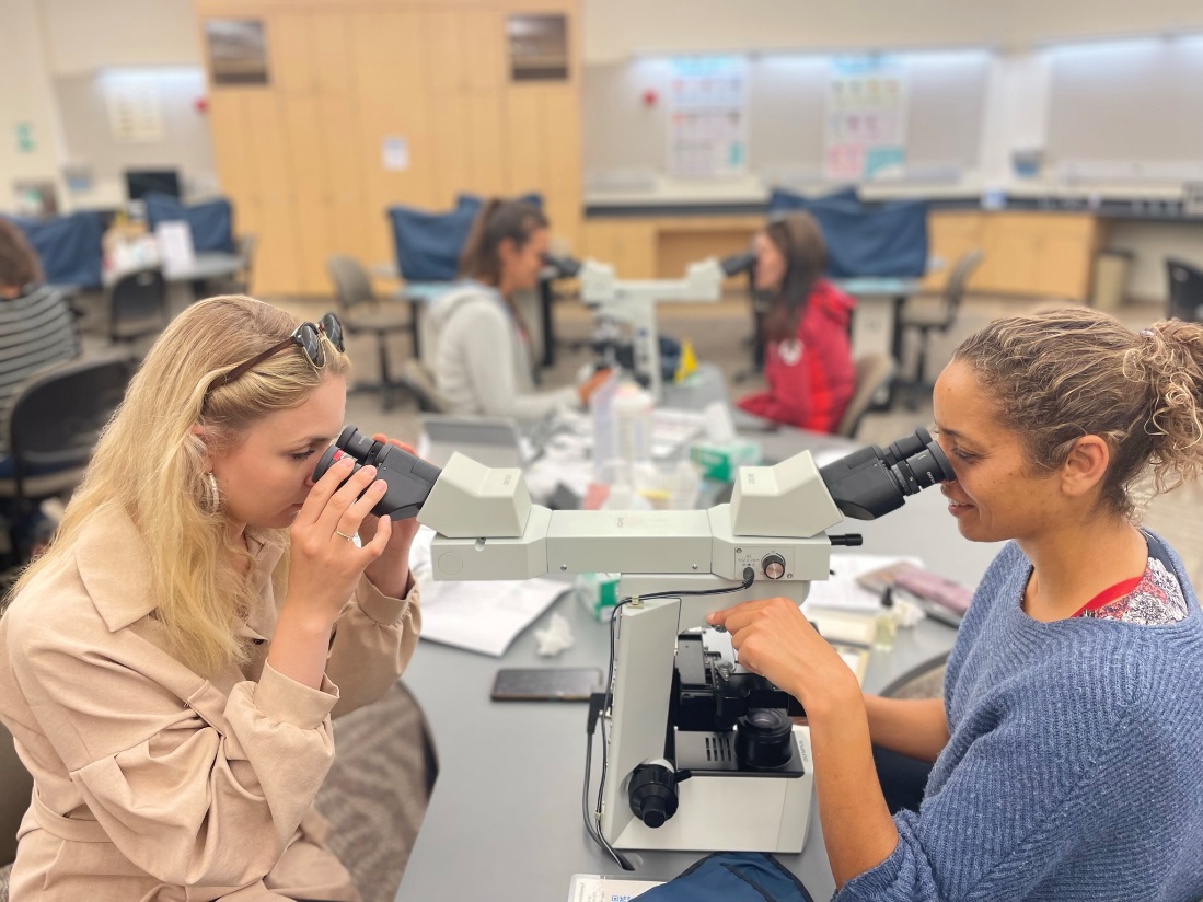 Evaluation of cytological preparations from material obtained from the conjunctival sac of companion and farm animals.
16th William Magrane Basic Science Course in Veterinary & Comparative Ophthalmology
Multi-Purpose Teaching Facility, UC Davis
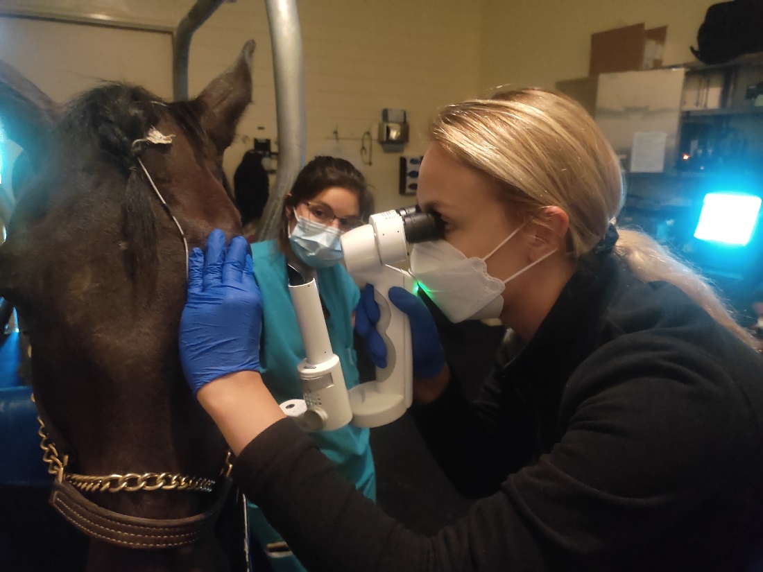 Ophthalmological examination of the horse using a slit lamp.
Uniwersytet Kalifornijski w Davis
School of Veterinary Medicine
Ophthalmology Service
The internship at the University of California allowed me to personally meet and make friends with scientists of the University of California, as well as lecturers of the course with whom I have started cooperation so far, incl. during the organization of webinars in the field of veterinary ophthalmology. The Ophthalmology Department is a group of people who are extremely open and willing to share their knowledge, putting cooperation in the team first and the only way to success in both scientific and clinical activities. The activities carried out so far and the possibility of an internship, as well as the help of the University of Life Sciences in Lublin, give the prospect of establishing further cooperation with the University of California, Davis.
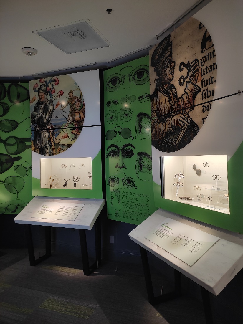 Museum of the Eye – American Academy of Ophthalmology
San Francisco, California, USA
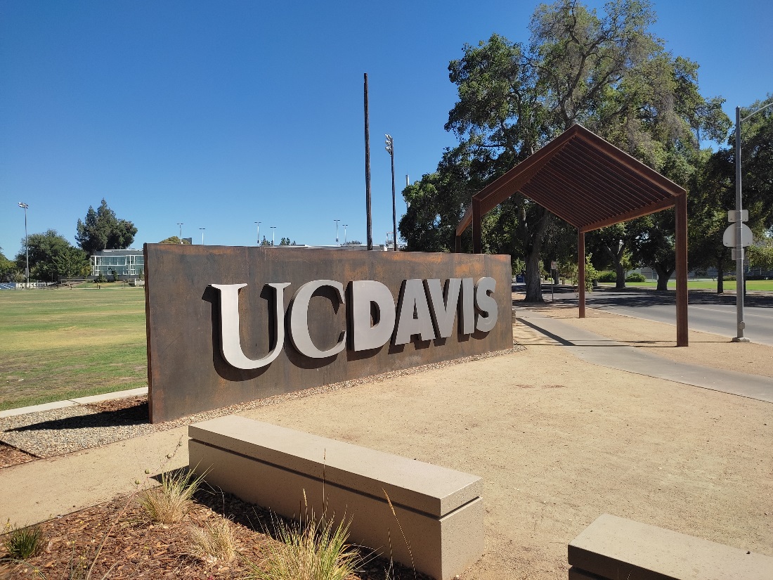 University campus
University of California, Davis
Internship tutors:
David J. Maggs, BVSc (hons), DACVO
Bianca Martins, DVM, MS, PhD; DACVO, DCLOVE (Hon)
Ed. Jowita Zwolska
13 Akademicka Street, 20-950 Lublin
VATIN 712 010 37 75
REGON no. 000001896
ePUAP: /UP-Lublin/SkrytkaESP